
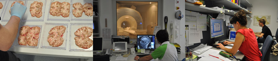

Neuromuscular Imaging
Research Outlook
Muscular dystrophies are a diverse group of genetic disorders that cause progressive muscle weakness and degeneration. Accurate monitoring of disease progression is essential for developing effective treatments. Our research unit explores advanced MRI techniques to assess metabolic and functional changes in patients with muscle disorders. By detecting early alterations in muscle tissue and correlating them with progressive fat infiltration, we aim to establish sensitive biomarkers for future therapeutic studies.
In addition to MRI analysis, we integrate clinical evaluations and other biomarkers, such as body fluids, to provide a comprehensive assessment of disease progression. Our research is conducted in close collaboration with neurologists and other research units at the Medical University of Graz, fostering a multidisciplinary approach to neuromuscular research.
Ongoing Studies
Updates coming soon.
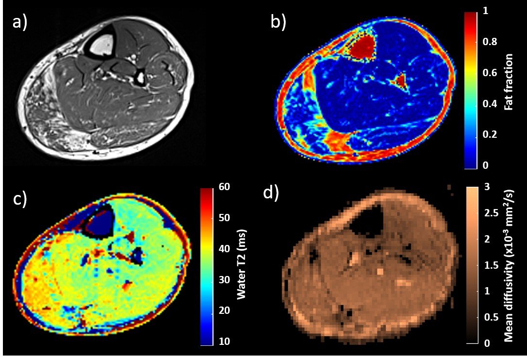
This figure shows MRI images of the lower leg from a patient with muscular dystrophy. Different MRI techniques allow us to visualize and analyze disease-related changes in muscle tissue:
- (a) Fat Replacement: Healthy muscle tissue is progressively replaced by fat, which can be detected and measured using MRI.
- (b) Fat Quantification: By analyzing MRI data, we can calculate the fat fraction in the muscle, providing an objective measure of disease progression.
- (c) Inflammation Detection: Some MRI techniques can reveal inflammation within the muscle, which may be an indicator of ongoing disease activity.
- (d) Structural Changes: MRI can also highlight alterations in muscle structure, helping to assess the overall integrity of the tissue.
By utilizing different MRI contrasts, we can obtain a comprehensive picture of disease progression, aiding in diagnosis, monitoring, and the evaluation of potential treatments.