
Postmortem MRI
Postmortem analyses of brain tissue offer the unique opportunity to improve our understanding of MRI contrast mechanisms by performing chemical and immunohistological correlation analyses. The frequently applied formalin fixation removes free water and consequently changes the magnetic susceptibility and MRI relaxation properties. Therefore, image contrast in fixed brain slices differs from the contrast acquired under in vivo conditions. To circumvent these limitations, we do post mortem analyses in fresh and unfixed brain tissue.
|
|
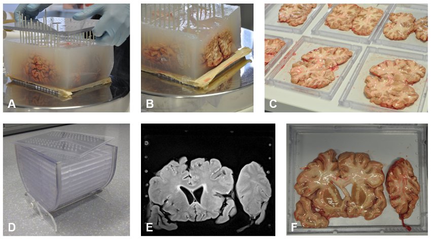
| Figure: Workflow of the postmortem study: The entire (unfixed) brain is embedded in agarose gel to maintain its structure and stiffness (A). Fiberglas rods arranged in 1cm spacing provide additional stability and enable precise cutting into 1 cm thick slices (B). Several Plexiglas trays holding the brain slices (C) are mounted inside the a plastic container which is filled with a buffer solution (D). The MRI (E) then is used as a reference to guide sample extraction for further histological and chemical analysis (F). |
Temperature mapping
Temperature mapping and correction of relaxivity changes induced by cooling of the tissue samples (fresh or in situ) are a prerequisite for quantitative MRI in postmortem tissue. In our current research, we assess the temperature-dependency of various relaxation parameters and develop correction schemes for different brain regions.
 |
| Figure 1: (a) Embedded and vacuumed brain slice. (b) Sealed sphere which enables controlled heating or cooling of the mounted brain slice by flushing with tempered water. (c) The refrigerated water bath with a heating immersion circulator. |
Traumatic brain injury (TBI)
Traumatic brain injury (TBI) is a major cause of death and disability, especially in children and young adults. Causes include falls, vehicle accidents, blunt trauma and violence. From a forensic point of view it is especially important to determine reliably whether the cause was an accident or abuse (e.g. shaken baby syndrome) as this information is a basis of jurisdiction in court.
The ultimate goal of this study is to improve the understanding of the extend of brain tissue injury when assessed by MRI. Those insights from postmortem analyses are translated to in-vivo condition and provide a basis for the forensic reconstruction of incidents.
| Figure: This study includes 20 subjects deceased of a traumatic brain injury (A) and 20 subjects deceased of non-traumatic reasons. Quantitative MRI takes place directly after death (B) . Following autopsy, several histologic (F,G) and chemical analyses (C,D) are carried out which provide in conjunction with the MRI an inproved understanding how fiber ruptures affect the MRI contrast (E, diffusion tensor imaging). |
References:
** Birkl, C., Langkammer, C., Haybaeck, J., Ernst, C., Stollberger, R., Fazekas, F. and Ropele, S. (2013)
Temperature-induced changes of magnetic resonance relaxation times in the human brain: A postmortem study.
Magn Reson Med. doi: 10.1002/mrm.24799
** Birkl, C., Langkammer, C., Haybaeck, J., Ernst, C., Stollberger, R., Fazekas, F., Ropele, S.
Temperature-induced changes of magnetic resonance relaxation times in the human brain: A postmortem study.
Magn Reson Med. 2013, doi: 10.1002/mrm.24799
** Langkammer, C; Krebs, N; Goessler, W; Scheurer, E; Yen, K; Fazekas, F; Ropele, S.
Susceptibility induced gray-white matter MRI contrast in the human brain.
Neuroimage. 2012; 59(2):1413-1419
** Langkammer, C; Schweser, F; Krebs, N; Deistung, A; Goessler, W; Scheurer, E; Sommer, K; Reishofer, G; Yen, K; Fazekas, F; Ropele, S; Reichenbach, JR
Quantitative susceptibility mapping (QSM) as a means to measure brain iron? A post mortem validation study.
Neuroimage. 2012; 62(3):1593-1599
** Langkammer, C; Krebs, N; Goessler, W; Scheurer, E; Ebner, F; Yen, K; Fazekas, F; Ropele, S
Quantitative MR imaging of brain iron: a postmortem validation study.
Radiology. 2010; 257(2):455-462